If you have diabetes mellitus, your body does not use and store glucose properly. Over time, diabetes can damage blood vessels in the retina, the nerve layer at the back of the eye that senses light and helps to send images to the brain. The damage to retinal vessels is referred to as diabetic retinopathy.
Nonproliferative Diabetic Retinopathy
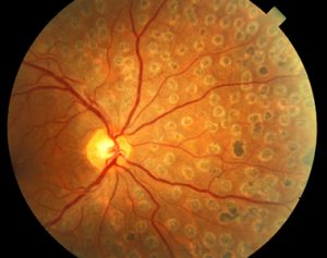
 Nonproliferative diabetic retinopathy (NPDR), commonly known as background retinopathy, is an early stage of diabetic retinopathy. In this stage, tiny blood vessels within the retina leak blood or fluid. The leaking fluid causes the retina to swell or to form deposits called exudates.
Many people with diabetes have mild NPDR, which usually does not affect their vision. When vision is affected, it is the result of macular edema or macular ischemia, or both.
Macular Edema
Macular edema is swelling or thickening of the macula, a small area in the center of the retina that allows us to see fine details clearly. The swelling is caused by fluid leaking from retinal blood vessels. It is the most common cause of visual loss in diabetes. Vision loss may be mild to severe, but even in the worst cases, peripheral (side) vision continues to function. Laser treatment can be used to help control vision loss from macular edema. Newer treatments are being investigated.
Macular ischemia occurs when small blood vessels (capillaries) close. Vision blurs because the macula no longer receives sufficient blood supply to work properly. Unfortunately, there are no effective treatments for macular ischemia.
Proliferative Diabetic Retinopathy
 Proliferative diabetic retinopathy (PDR) is a complication of diabetes caused by changes in the blood vessels of the eye. If you have diabetes, your body does not use and store sugar properly. High blood sugar levels create changes in the veins, arteries, and capillaries that carry blood throughout the body. This includes the tiny blood vessels in the retina, the light-sensitive nerve layer in the back of the eye.
In PDR, the retinal blood vessels are so damaged they close off. In response, the retina grows new, fragile blood vessels. Unfortunately, these new blood vessels are abnormal and grow on the surface of the retina, so they do not resupply the retina with blood.
Occasionally, these new blood vessels bleed and cause a vitreous hemorrhage. Blood in the vitreous, the clear gel-like substance that fills the inside of the eye, blocks light rays from reaching the retina. A small amount of blood will cause dark floaters, while a large hemorrhage might block all vision, leaving only light and dark perception.
The new blood vessels can also cause scar tissue to grow. The scar tissue shrinks, wrinkling and pulling on the retina and distorting vision. If the pulling is severe, the macula may detach from its normal position and cause vision loss.
 Laser surgery may be used to shrink the abnormal blood vessels and reduce the risk of bleeding. The body will usually absorb blood from a vitreous hemorrhage, but that can take days, months, or even years. If the vitreous hemorrhage does not clear within a reasonable time, or if a retinal detachment is detected, an operation called a vitrectomy can be performed. During a vitrectomy, the eye surgeon removes the hemorrhage and any scar tissue that has developed, and performs laser treatment to prevent new abnormal vessel growth.
People with PDR sometimes have no symptoms until it is too late to treat them. The retina may be badly injured before there is any change in vision. There is considerable evidence to suggest that rigorous control of blood sugar decreases the chance of developing serious proliferative diabetic retinopathy.
 A medical eye examination is the only way to discover any changes inside your eye. If your ophthalmologist or optometrist finds diabetic retinopathy, you may require a special test called fluorescein angiography or optical coherence tomography (OCT) to find out if you need treatment.
If you have diabetes, early detection of diabetic retinopathy is the best protection against loss of vision. You can significantly lower your risk of vision loss by maintaining strict control of your blood glucose and visiting your ophthalmologist regularly. People with diabetes should schedule examinations at least once a year. Pregnant women with diabetes should schedule an appointment in their first trimester, because retinopathy can progress quickly during pregnancy. More frequent medical eye examinations may be necessary after a diagnosis of diabetic retinopathy.